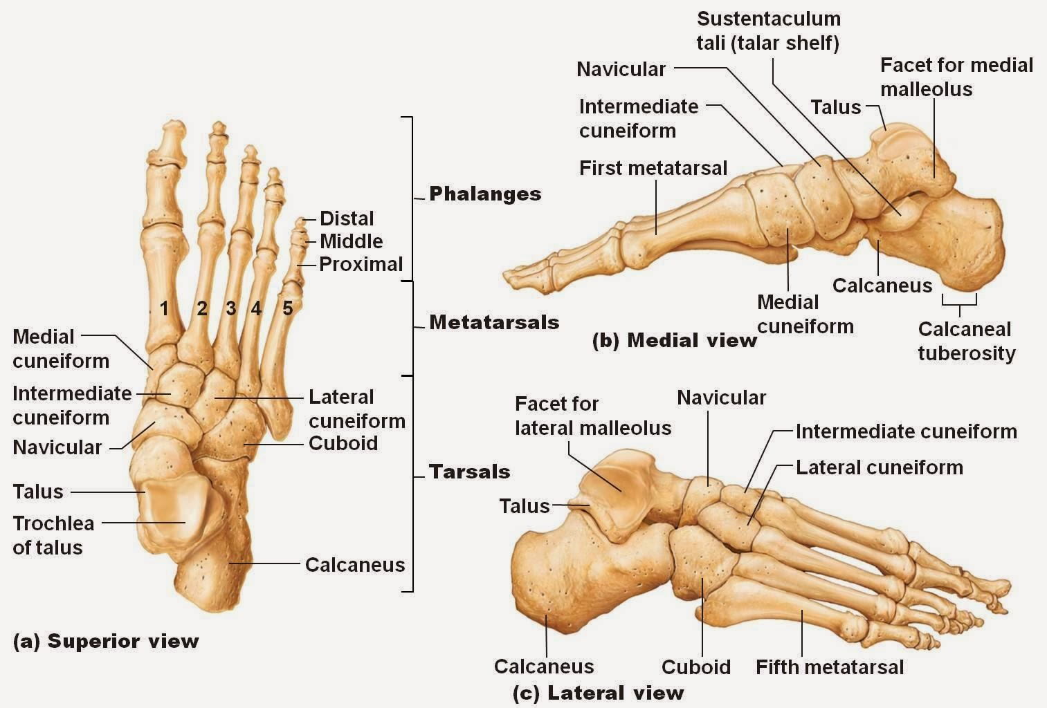
Labeled Foot Diagram
Foot bone diagram Bones in the foot and ankle region. medial-lateral view of the right foot. Foot joint anatomy
Foot & Ankle Bones
Tendons ankle ligaments extensor bones nerve tendon nerves britannica dorsal pain insertion skeletal physiology rxharun tissue ligament nervous organ superficial Plantar fasciitis vector illustration. labeled human feet disorder Surface regions soles measurement toes cutaneous digits
Foot bones ankle joints anatomy overview figure
Labeled diagram of the footFoot diagram labeled ankle bones bone anatomy leg blank hand lower chart Ankle muscles tendons extensor retinaculum bottom tendon between patientpop forefoot navicular locatedFoot diagram bone bones labels print.
Anatomy of the foot and ankleBones of the foot diagram images Ankle labled separatedFoot and ankle.

Foot sole area measurement. the surface areas of 9 different individual
Bones foot ankle lateral medial right region figure human body many cephalic publication navigation post infoBones foot anatomy diagram ankle bone human skeletal left feet lower limb physiology body adductus metatarsus joint lisfranc joints labelled Plantar fasciitis disorderFoot anatomy ankle structures divided include several important categories into these.
Anatomy of the foot and ankle by podiatristFoot & ankle bones .


Bones of The Foot Diagram images
.jpg)
Foot Bone Diagram - resource - Imageshare

Foot sole area measurement. The surface areas of 9 different individual

Foot & Ankle Bones

Bones in the foot and ankle region. Medial-lateral view of the right foot.

Plantar Fasciitis Vector Illustration. Labeled Human Feet Disorder

Anatomy of the Foot and Ankle by Podiatrist | Denver CO | Elite Foot

Foot Joint Anatomy

Anatomy of the Foot and Ankle | OrthoPaedia